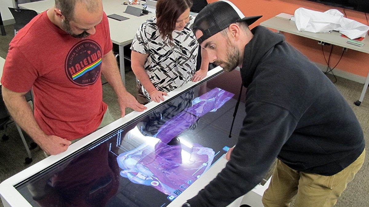
Four human bodies inside a high-tech table at the Allan Price Science Commons and Research Library are drawing the attention of University of Oregon students studying anatomy.
“This table is the next best thing to having a real human cadaver body to study,” said Jon Runyeon, a senior instructor in the Department of Human Physiology and director of the human anatomy laboratory.
The $80,000 table — made by Anatomage and billed as the world’s first virtual dissection table — was gifted to the department last fall by UO alumnus Samuel Blackwell, a retired Redmond physical therapist, and his wife, Linda. The table was installed in December into the human physiology tutorial room in the lower level of the science library.
Looking through the clear top, users can access the complete bodies of the donors whose digitized remains were loaded slide by slide after dissection. Their reconstruction provides accurate 3D views. With fingertip touches on the screen, users can zoom into and change angles to study a body part and learn how components are interconnected.
Since winter quarter started, the table has been used by anatomy students as a complementary way to dive into the details they only can glimpse during limited class time in the anatomy laboratory, also known as the cadaver dissection lab. The tutorial room is open during library hours, giving easy access each quarter to some 300 students.
“It’s a really powerful tool for looking at the anatomy of a human body,” Runyeon said. “The primary difference between using the table and using a real cadaver is the kinesthetic difference. This tool is more visual.”
The bodies are unique on both the outside and inside, he said.
“Students can learn about these different anatomical variations,” Runyeon said. “This can be especially important for future surgeons who need to realize such differences.”
The table also contains reference databases of various body components, including more than a thousand diseased states.
While demonstrating the possibilities, Runyeon touched the screen to unveil a brain’s temporal and frontal lobes. As he zoomed in, the gyri, or ridges, were easily seen, along with bundles of myelinated axons, or white matter.
What is seen on the table can be projected to a monitor. The table also can be stood on end. Views can be manipulated to show body components with or without icons, and users can write, add text or draw arrows on the screen as they explore. There are options for playing games and taking quizzes.
“It’s like a giant iPad,” said Angie Towle, an accounting technician and social media coordinator in the human physiology department. Students love it, she added.
“What’s really fun is being able to go over structures with other students,” said Mike Stacey, a junior who is double majoring in human physiology and psychology who was studying in the tutorial room during the demonstration. “The level of detail is really remarkable.
“There is a game mode that’s really fun,” he said. “It incorporates the excitement of new technology and learning. It provides different methods of learning different structures. In the game mode you can have four people around the table and play Jeopardy. You buzz in if you can identify something. If a person is wrong, the next person can try.”
Stacey, who graduated from South Eugene High School and moved to Vancouver, British Columbia, to attend paramedic school, is now planning to pursue medical school.
For now, Runyeon said, the table is being used for additional study time. Eventually, he said, it may be used as part of course assignments. Requests for access to the table, he added, are coming in from faculty members across campus who believe their courses might benefit from using the cadaver table.
—By Jim Barlow, University Communications


