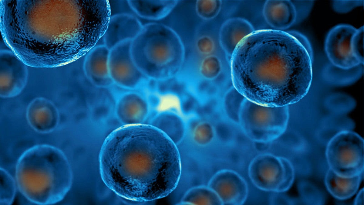
What seems like a straightforward task for a cell — dividing in two — is actually an intricate series of engineering puzzles. A dividing cell needs to maneuver its insides so that the right components will end up in each cell, to produce two functioning cells.
University of Oregon biochemist Ken Prehoda is trying to solve one of those fundamental puzzles: how a dividing stem cell portions out its membrane during the process of division.
In a new study, he and postdoctoral researcher Bryce LaFoya show how ready-to-divide stem cells create a reservoir of extra membrane, which accommodates the increased surface area necessary for two cells. The pair describe their findings in a paper published April 27 in Developmental Cell.
Animal cells are surrounded by a thin membrane that forms a protective barrier around the cell. Just before an animal cell divides, it gets rounder, said Prehoda, who is part of the College of Arts and Sciences.
“Geometrically, a sphere is optimal for minimizing the amount of membrane,” he said. “But to divide the cell in two, it gets squeezed and the surface area sharply increases.”
In that sense, cell division is like pinching a balloon, except that balloons can stretch as they change shape. A cell’s membrane isn’t stretchy like a balloon’s, but it still needs to be able to expand somehow to accommodate squeezing together and pinching off a new cell.
Prehoda’s team focused on this challenge in neural stem cells, which are the cells that generate the cells in our nervous system. Because they keep churning out new cells, stem cells divide asymmetrically: The stem cell holds onto most of the cell’s material, giving just a little bit away to the new sister cell.
Prehoda and LaFoya used a spinning disk confocal microscope equipped with cutting-edge super-resolution technology to peer inside the brains of developing fruit flies. LaFoya noticed that neural stem cell membranes were decorated with folds and small protrusions, “kind of like the extra skin on wrinkly dog breeds like shar-pei and bulldogs,” he said.
LaFoya realized that the neural stem cell membrane uses these “wrinkles” to save up membrane for when it’s needed during division. As LaFoya watched the cells divide, he was amazed to see the wrinkles collect on one side immediately before division. The positioning of the wrinkles determined where the cell could grow and where the new cell could form.
While dividing unequally allows the neural stem cell to only give up a fraction of its resources, the asymmetry makes partitioning the membrane an especially tricky problem. LaFoya noted that the cell has found a remarkable solution to that part of the engineering puzzle.
“Making the extra membrane beforehand and positioning it properly before division is an elegant solution that we hadn’t envisioned before starting this project,” he said.
That is just one of many questions Prehoda and his lab are trying to answer with advanced microscopy techniques, which allow them to look at cells in new levels of detail. Thanks to this new technology, Prehoda said, “we have an embarrassment of riches, and lots of new imagery to explore.”
—By Laurel Hamers, University Communications
This research was funded by the National Institutes of Health.


