The mysteries of teenage brains. The genetic underpinnings of schizophrenia. How we take in a friend's face with our eyes and commit it to memory. Are we closer to reeling in such understanding?
UO scientists say their recent project, soon to be detailed in the Journal of Neurophysiology, puts them firmly on the path of the White House BRAIN Initiative. The initiative, known as the Brain Research through Advancing Innovative Neurotechnologies, seeks to understand how brains process sights, sounds and other input and turn that into actions and responses.
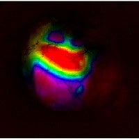
In the research, led by UO doctoral student Joseph B. Wekselblatt, a team developed a brain-visualization technique that allows the use of living mice to explore areas of the brain that functional MRI scans of humans can only highlight generally. The approach captures neurons as they activate in real time, during the processing of sensory stimuli through subsequent behavioral responses.
Human brain studies done with fMRI — a specialized form of magnetic resonance imaging — allows researchers to pinpoint regions of the brain that are active under certain conditions by measuring changes in blood oxygen levels. It does not allow researchers to see the specific neuron circuitry.
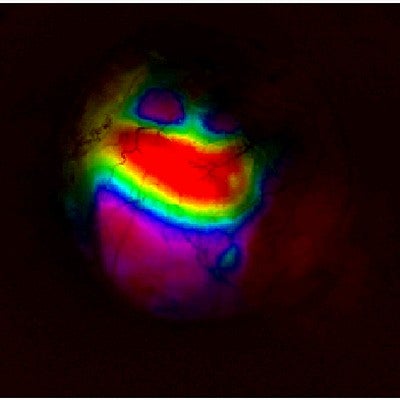
"Our technique is like fMRI but with far greater temporal and spatial resolution," said Cristopher M. Niell, a professor in the Department of Biology and member of the Institute of Neuroscience. "We can visualize sensory inputs as they come into the brain, and the subsequent activity corresponding to a decision and behavioral response. We see the whole flow."
The idea, Niell said, is that human brain regions identified by fMRI can be looked at more closely in the simpler mouse model to explore basic mechanisms.
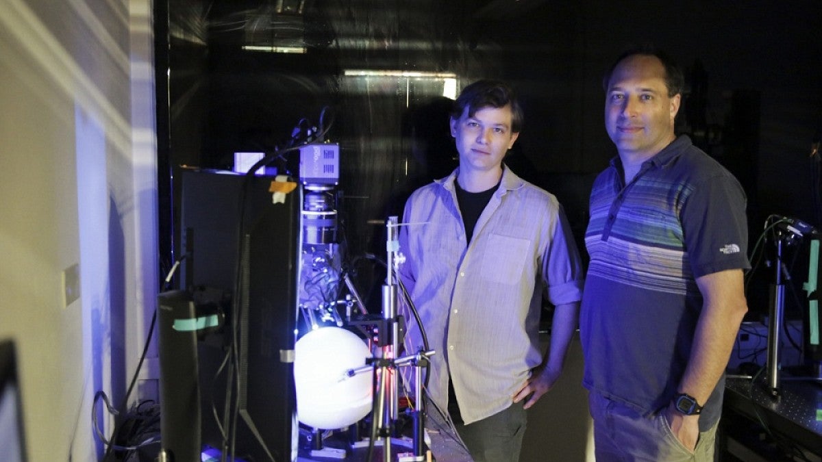
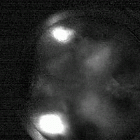
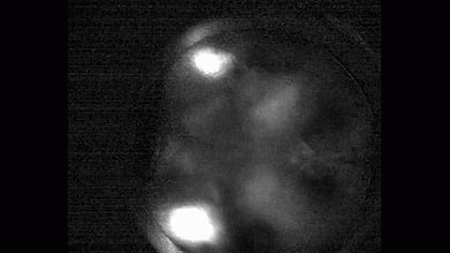
For the technique, UO researchers developed a line of transgenic mice whose brains contain a green fluorescent protein that lights up active neurons. They then used a customized wide-field microscope with dual lenses to capture images of the brain similar to what fMRI does in humans. The final step involves two-photon imaging, which zooms in and locates neurons that are active. Combined, they see activity across the cortex, the outer surface of the brain where cognition, memory, language learning and motor behaviors occur.
The fluorescent protein used by the UO team was developed at the Howard Hughes Medical Institute. It contains a calcium sensor and lights up when neurons are activated. The mouse line generated at the UO is now available to scientists around the world through the Jackson Laboratory in Maine.
"We deliver sensory inputs — moving images — that trigger decision-making by the mouse," Niell said. "As the inputs are registered and behavior begins, we can watch the flow of activity across the brain. You see it all in real time, and very quickly, nearly at the speed of thought."
"In previous research," Wekselblatt said, "you'd have to use different animals at different times of their lives to get to information that you want. Here we can study the same mice over time to observe how patterns change when they are exposed to different variables, such as stress or medications."
Niell's lab already is exploring how the technique might help identify genes associated with schizophrenia. Plans also are underway to work with researchers in the UO Department of Psychology and College of Education who are studying adolescent brain activity, ranging from basic development to risky behaviors. Researchers plan to explore the impacts of interventions on the brain and whether some teens are more brain-ready than others for interventions that seek to improve such things as planning, decision-making and setting goals. Another possibility for the technique, Niell said, is to look at age-related deterioration of the brain.
Co-authors with Niell and Wekselblatt are Erik D. Flister, a doctoral student, and Denise M. Piscopo, a staff scientist in Niell's lab. The National Institutes of Health and the Searle Foundation supported the research.
—By Jim Barlow, University Communications


