Zebrafish Reveal How Bioelectricity Shapes Muscle Development
New research builds on work University of Oregon neuroscientists started 40 years ago
A question left unanswered in a biologist’s lab notebook for 40 years has finally been explained, thanks to a little fish that couldn’t wriggle its tail.
New research from the University of Oregon describes how nerve cells and muscle cells communicate through electrical signals during development—a phenomenon known as bioelectricity.
The communication, which takes place via specialized channels between cells, is vital for proper development and behavior. The study identifies specific genes that control the process, and pins down what happens when it goes wrong.
The finding offers clues to the genetic origins of muscle disorders in humans, and taps into longstanding questions in developmental biology.
“This is something many of us have wondered about for many, many years—and now we’ve figured it out!” said Judith Eisen, the UO neuroscientist who, in the 1980s, spotted a communication pattern between zebrafish muscle cells that she couldn’t explain.
Eisen and her colleagues report their findings in a paper published June 26 in Current Biology.
The work ties together three generations of UO neuroscientists and provides a lesson for all researchers: keep those lab notebooks. Eisen unearthed her original hardcover notebooks when moving into temporary lab space for a building renovation a few years ago. The sketches and shorthand notes she recorded in ink years ago are still relevant today.

A Muscular Mystery
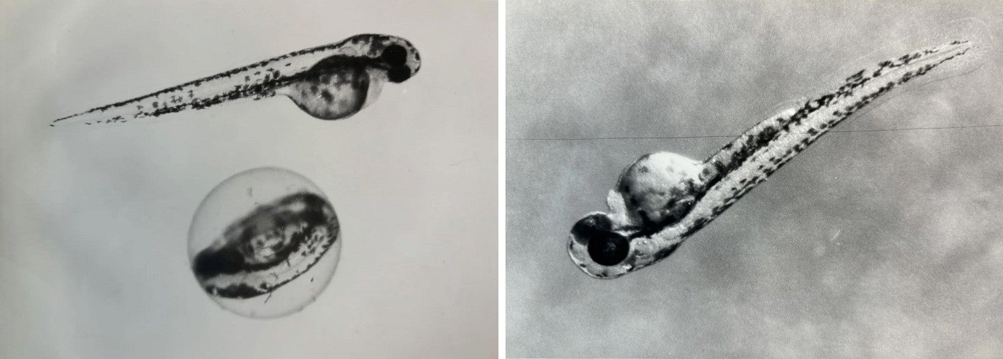
In 1983, Eisen was a postdoctoral researcher in the lab of Monte Westerfield, just beginning her career at the UO. She was part of a small group of scientists working to establish zebrafish as a new model organism, hoping to use these small, shimmery fish to probe questions about the development of vertebrate animals.

Model organisms like mice, fruit flies and worms allow scientists to do experiments that aren’t possible in humans, answering fundamental biological questions and providing guidance for more focused testing in humans.
Zebrafish were a promising addition to the scene. Zebrafish and humans share many genes, making the fish useful for testing the genetic underpinnings of human diseases and conditions. And because zebrafish embryos are transparent, scientists can watch development happen in real time under the microscope.

But at the time, everything about this system was new. Biologists had to figure out how to care for the fish in the lab and effectively use them in experiments.
One day, Eisen and Westerfield were using a yellow tracing dye to highlight individual nerve cells in the zebrafish, for observation under the microscope. The cells they wanted to reach could only be accessed by inserting a pipette filled with the glowing dye through the muscles. So some dye ended up mingling with the muscle cells, too.
Eisen and Westerfield were intrigued by the way the dye spread through the muscle cells. It spread cell-to-cell in a way that suggested that the cells were sharing messages directly—via some physical connecting channel between them, rather than via longer-range chemical messengers.
This didn’t fit the understanding of how adult muscle cells communicate with each other. But there was a dawning realization in the field that connections between muscle cells might be important during muscle development.
Eisen sketched out what she saw in her lab notebook, as did Westerfield. But there wasn’t a good way to probe further, Eisen said. While scientists at the time knew these sorts of communication channels existed, they didn’t know the genes that created them, or have the tools to ask what they were doing. So it was a dead end.
Eisen moved onto other questions, making major contributions to the field of developmental biology over the course of her career. This April, she was inducted into the National Academy of Sciences, one of the most prestigious honors for a scientist.
Over the last 40 years, Eisen and her UO colleagues, alongside scientists around the world, have continued to develop the zebrafish as a model organism. The advance of genetic technologies has made this little fish an even more powerful ally for understanding biology.

A Fish That Couldn’t Swim
A few years ago, Eisen’s observation resurfaced in the lab of another UO neuroscientist, Adam Miller.
Miller was recruited to the University of Oregon by Eisen and colleagues to build a research group focused on electrical communication between cells. His lab studies how neural circuits build connections and create behavior. One area of focus is gap junctions, physical channels that allow electrical signals to move directly between cells. These communication pathways are particularly important during early development, as the body’s many systems are getting set up and organized.
Zebrafish are the perfect species to study electrical communication. Thanks to their transparent embryos, “we can image electricity flowing through cells in real time,” said Rachel Lukowicz-Bedford, a postdoc in Miller’s lab.
While hunting for zebrafish with different gap junction mutations, Lukowicz-Bedford made an intriguing find: a zebrafish that couldn’t move its tail properly. Usually, a zebrafish embryo will flop around and spontaneously flick its tail, but these fish didn’t do that.
As they did experiments to figure out why, the team realized this fish might be a possible link back to Eisen’s observation in muscle cells in the 1980s.
This was a really cool University of Oregon zebrafish project because [Eisen and Westerfield] knew that this existed for a really long time, but at the time they were originally doing these experiments, they didn't have ways address the complex genetic landscape that we do now," Lukowicz-Bedford said. "I don't think these experiments would have come together anywhere else.
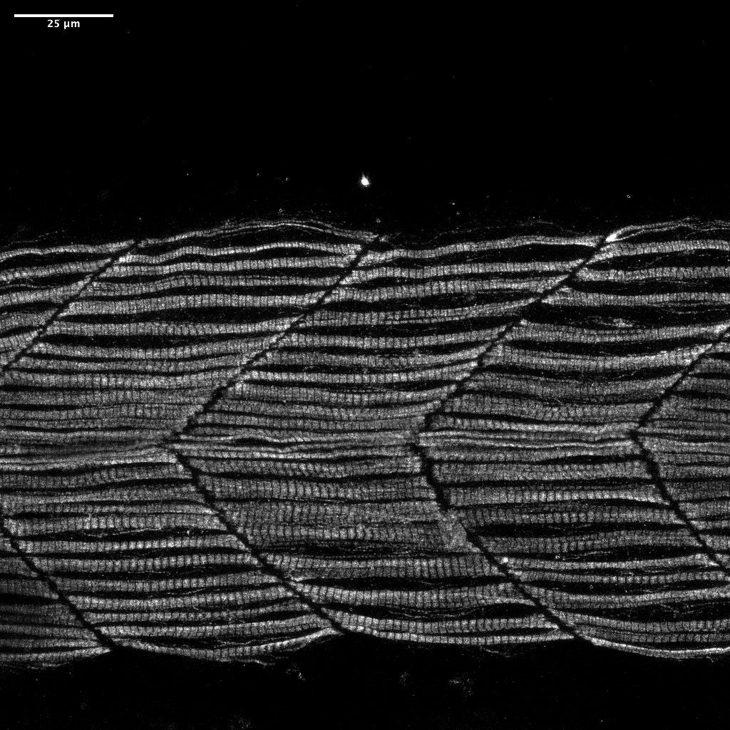
In healthy zebrafish, researchers can watch the electrical signals propagate through the gap junctions between muscle cells, like a plume of food dye diffusing into a cup of water. In fish with this mutation, the signals don’t flow. The mutation was impairing electrical communication between the cells via the gap junctions.
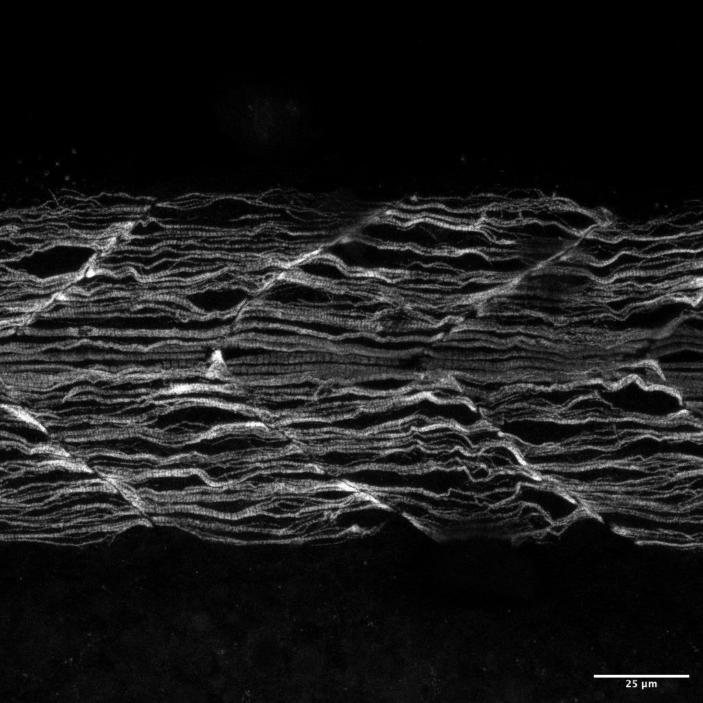
And that communication breakdown led to improper muscular development, they showed. In an ordinary healthy zebrafish, the muscle fibers are straight and orderly. In the zebrafish with this mutation, the muscle fibers are crinkly and wavy, like crepe paper streamers.


The researchers pinned the change on a mutation in a specific gene. Through a series of experiments, they showed that this gene, when functioning normally, makes the gap junction channels between muscle cells that allow the nervous system to coordinate the activity of early developing muscle. And without appropriate electrical signaling at the right time during development, the muscle fibers can’t organize properly, causing crinkly muscle fibers and severe muscle defects.
“We figured out that this gap junction channel is a conduit—it allows electricity from the nerve cells to be sent out to muscle fibers,” Lukowicz-Bedford said.
The finding answers Eisen’s decades-old question, sketched out in a lab notebook that she still has: The yellow dye was moving between muscle cells because of these specific communication channels.
More than a curiosity, though, the findings can help inform scientists’ understanding of muscle development in humans. In disorders where muscles don’t develop properly, faulty gap junction channels might be one cause, a link that was previously unknown.
“The gene we studied in this paper is not a weird zebrafish gene; it’s also found in humans,” Lukowicz-Bedford said. “By using zebrafish, we can go after this gene with basically unknown function in humans, and be able to understand what it’s doing in context. We’ve been able to uncover the function of a gene that’s been really elusive.”
The research also illustrates that electrical signaling between different systems is critical for development. Similar communication is probably at play in the development of other body systems, too, the researchers suggest—it’s likely not specific just to muscles.
“The transfer of bioelectricity from one organ system to another is critical for development and adult function,” Miller said. “Finding the genes that allow this to occur, understanding how they work, and exactly what goes wrong when communication is disrupted, will provide new insight into human disease.”
This research was funded by the National Institutes of Health.
Zebrafish International Resource Center
The University of Oregon is home to the Zebrafish International Resource Center, which shares different genetic varieties of zebrafish with researchers around the world.

